
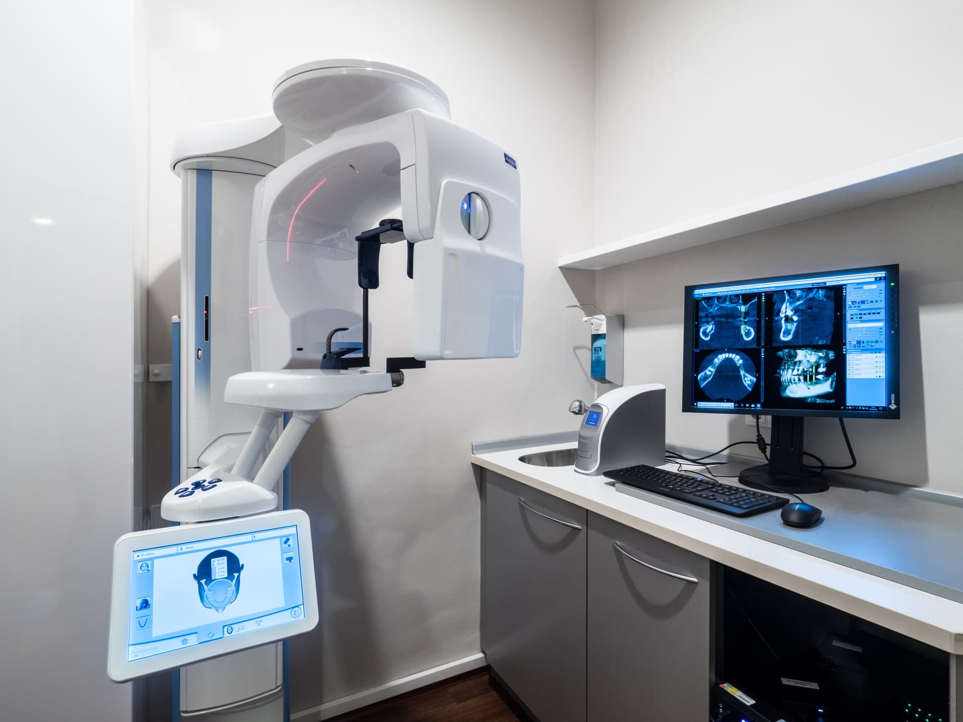
3D X-ray (CBCT)
Cone beam computed tomography (CBCT – known in German as DVT) produces three-dimensional X-ray images.
It provides cross-sectional images in all three planes, similar to computed tomography (CT). Teeth and jawbone are shown in their exact spatial position. The radiation dose is higher than for a conventional two-dimensional X-ray; how much higher depends on the device and the size of the area scanned.
Depending on the clinical question, this can offer patients and dentists the following advantages:
- A three-dimensional basis for diagnosis and planning: neighbouring structures such as the maxillary sinuses and nerve canals can be located precisely and taken into account.
- Changes that may be causing symptoms, such as cysts, can be detected and located precisely, even where they remain hidden on a conventional 2D X-ray.
- In implant dentistry in particular, the scan supports the planning of the procedure.
- A three-dimensional view on screen illustrates the planned treatment and makes it easier for patients to understand.
- The scan is taken while you stand or sit – there is no need to lie in a confined tube.
CBCT is generally not covered by statutory health insurance and is billed privately. Whether, and to what extent, private health insurance or supplementary insurance covers the costs depends on your policy.
Standard X-ray diagnostics are, of course, still available: two-dimensional X-rays (single-tooth and panoramic) in good image quality, keeping the radiation dose as low as reasonably achievable. We decide which type of image makes sense in each case based on your findings.